USG Lab
Reception:
In person: 8:00 am to 7:00 pm
By phone:
National Health Fund Visits +48 12 413 00 99
Paid Fund +48 606 445 411
The following data is required to make an appointment:
- Full address with postal code,
- Patient PESEL [ID] number.
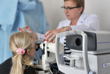
USG – cross-sectional images of examined organs
We operate modern ultrasound equipment. The results can be provided to you in print or digital format, and are always accompanied by the analysis of the investigating doctor.
We provide the USG imaging services:
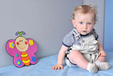
- Pelvic ultrasound (children),
- Ultrasound of gastrointestinal tract and abdomen (children),
- Neonatal cranial ultrasound scan (children),
- Hip ultrasound (children),
- Ultrasound of salivary glands (children and adolescent),
- Thyroid ultrasound (children and adolescent),
- Ultrasound of urinary system (children),
- Ultrasound of lymph nodes (children),
- Abdominal ultrasound (children),
- Other (children).
The ultrasound scan is safe, non-invasive and painless. It uses high frequency acoustic waves that are not audible to the human ear. Therese waves penetrate the body and dissolve without damaging the cells. The reflected waves create an image visible on the screen.
Preparation for ultrasound
Reception will inform you how to prepare for your particular ultrasound.
Here are some specific recommendations:
- Newborns and infants – no preparation required. It is recommended that you feed or provide a drink to your baby beforehand. Mothers of infants should bring a bottle of food or drink with them to the appointment.
- Abdominal ultrasound in older children are carried out 3–6 hours after a meal at the earliest. Two days before their ultrasound, older children should follow a light diet, without carbonated beverages, and take Espumisan, as directed by the referring doctor.
- Every patient visiting for an abdominal or pelvic ultrasound examination should have a full bladder. This can be achieved by drinking plenty of still water before (30–60 minutes) ultrasound.
Ultrasound
The doctor applies a gel to the skin, which facilitates the conduction and eliminates air bubbles before applying the scanner head. The gel is indifferent to the skin and easily removable. Moving the scanner head around, the doctor obtains a complete picture of the organ to be examined.
 Specialist clinics
 Neonatal Care Clinic